#Bizwhiznetwork.com Innovation ΛI |Technology News
A state-of-the-art CT scanning technology has shed fresh light on Megalosaurus bucklandii, the first dinosaur ever named and described scientifically — thanks to new research from the Universities of Warwick and Oxford, UK.

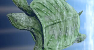
Artist’s impression of how Victorian paleontologists thought Megalosaurus bucklandii looked (right), compared with how we now understand it to have looked (left). Image credit: Mark Garlick / University of Warwick.
Megalosaurus bucklandii was a carnivorous dinosaur that lived in the Middle Jurassic, around 167 million years ago. It would have been about 30 feet (9 m) long and weighed about 1,400 kg.
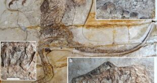
The world-famous Megalosaurus jawbone, collected over 200 years ago in a quarry near the village of Stonesfield in Oxfordshire, UK, is housed at the Oxford University Museum of Natural History.
Using the X-Ray Computed Tomography (XCT) and specialist 3D analysis software, Professor Mark Williams of the University of Warwick and co-authors took more than 3,000 X-ray images of the jawbone, creating a digital 3D image of the specimen.
In an unprecedented level of analysis, they were able to see inside the jawbone for the first time, tracing the roots of teeth and the extent of different repairs.
Some damage occurred to the specimen when it was removed from the rock, possibly shortly after it was discovered.
Records at the Oxford University Museum of Natural History suggest that some restoration work may have been undertaken by a museum assistant between 1927 and 1931, while repairing the specimen for display.

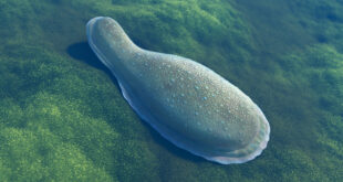
The jawbone of Megalosaurus bucklandii and 3D printed replica. Scale bars – 5 cm. Image credit: Wilson et al.
The XCT scans have revealed previously unseen teeth that were growing deep within the jaw before the animal died — including the remains of old, worn teeth and also tiny newly growing teeth.
The scans also show the true extent of repairs on the fossil for the first time, revealing that there may have been at least two phases of repair, using different types of plaster.
“Being able to use state-of-the-art technology normally reserved for aerospace and automotive engineering to scan such a rare and iconic natural history specimen was a fantastic opportunity,” Prof. Williams said.
“When I was growing up I was fascinated with dinosaurs and clearly remember seeing pictures of the Megalosaurus jaw in books that I read.”
“Having access to and scanning the real thing was an incredible experience.”
The research was presented on May 25, 2017 at the Institute of Electrical and Electronics Engineers (IEEE)’s International Instrumentation and Measurement Technology Conference in Torino, Italy.
_____
Paul Wilson et al. 2017. Utilizing X-Ray Computed Tomography for heritage conservation : the case of Megalosaurus bucklandii. In: I2MTC 2017 IEEE International Instrumentation and Measurement Technology Conference